
We believe that sharing data, software and research tools helps accelerate scientific progress and clinical innovation. Our open research resources support collaboration, transparency and reproducibility across the medical imaging and radiation therapy community.
Open source
Through GitHub, we share selected code repositories, open-source tools, research software and technical resources developed at Image X Institute.
View our open source resources here.

Public data
Our collections of anonymised clinical data are freely available for research purposes. Please acknowledge the data source via citation for any publications or presentations that use this data.
Tools for benchmarking real-time radiotherapy systems
Included in this data are lung [1] and prostate [2] patient measured motion traces, CT and contour sets (defined as per RTOG 1021 and RTOG 0938 respectively).
Robotic QA Phantom 6DoF
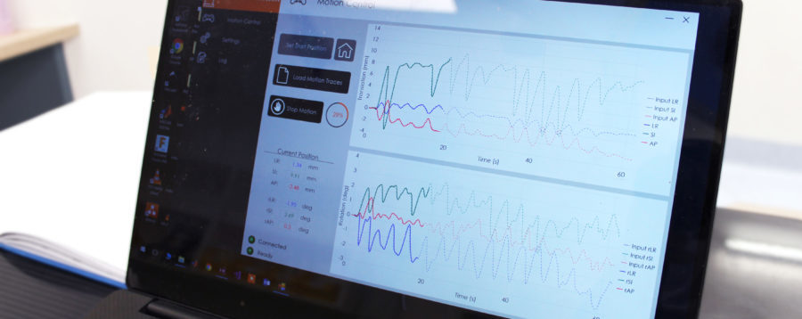
The robotic phantom quality assurance device accurately mimics tumour motion, ensuring safe implementation of radiotherapy technologies in the clinic. This project is now available open source – we have provided the software, as well as specifications for equipment needed, and full documentation.
TROG 15.01 Stereotactic Prostate Adaptive Radiotherapy utilizing Kilovoltage Intrafraction Monitoring (SPARK) Database
This dataset contains 48 prostate cancer patient data including the planning CT images, treatment plans, RT structure sets, planned and motion-included dose volume histograms, intra-treatment kilovoltage (kV) and megavoltage (MV) projection images, tumor translational and rotational motion determined by KIM, tumor motion ground truth data determined by the triangulation method, the linear accelerator trajectory traces and patient treatment outcomes.
The Sparse-view Reconstruction Challenge for Four-dimensional Cone-beam CT Dataset
The goal of the SPARE challenge was to explore the possibility of high quality 4D CBCT while sparing patients the additional scan time and imaging dose that was currently used in the clinics. 4D CBCT allowed the verification of tumor motion for thoracic patients immediately before every radiotherapy session. A clinical 4D CBCT scan took 2–4 minutes, as compared to one minute for a 3D CBCT scan. Participants received CBCT projection data either acquired clinically or simulated. Prior CT data and training sets were also be provided. Using these datasets, participants applied their reconstruction algorithms and submitted the final reconstructions to the host within a timeframe of three months.
4D-Lung
This data collection consists of images acquired during chemoradiotherapy of 20 locally-advanced, non-small cell lung cancer patients. Contribution from Prof Paul Keall prior to the creation of the Image X Institute.
Image X GitHub
Explore our repositories and open-source resources.