News
March 2025
NHMRC Grant Success for Tess Reynolds and Paul Keall
Both Paul Keall and Tess Reynolds have been awarded highly competitive NHMRC Investigator Grants. These are 5-year grants that support the investigators and provide a research support package. As is Image X tradition, we have celebrated with sugar – the team were treated to gelatos on the afternoon the good news came in.
Paul Keall, awarded $3M – Advancing image guided radiation therapy from discovery to clinical practiceIn this Investigator Grant I will dramatically improve cancer treatment outcomes by advancing a pipeline of image guided radiation therapy innovations from discovery to clinical practice. As radiation therapy is recommended for half of the 20 million cases of cancer diagnosed annually around the world, advanced radiation therapy innovations from this grant will impact over 10 million cancer patients every year.
Tess Reynolds, awarded $1.6M – Unlocking the future of robotic imaging for surgical procedural navigation and verification.
Robotic imaging systems have revolutionised surgical interventions.
To deliver the future of 3D image guidance for challenging surgical interventions, where options remain limited, I will develop imaging technologies that promise to allow direct in-room surgical verification of orthopaedic hardware, adaptively sync to the patient to mitigate motion without mechanical intervention and facilitate long anatomical sites to be captured in a single image to assess anatomy alignment intraoperatively.
Oct 2024
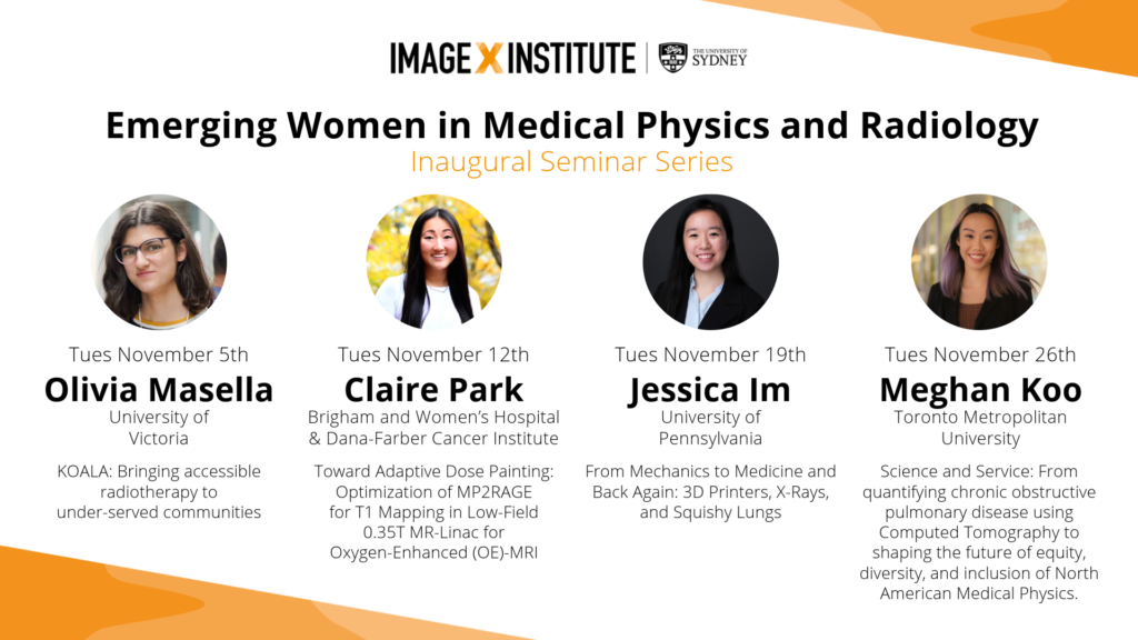
Inaugural Emerging Women in Medical Physics and Radiology Seminar Series
This November we’ll be holding our first inaugural Emerging Women in Medical Physics and Radiology Seminar Series. Join us on Tuesdays at 11am on zoom to learn about the amazing work being done across a range of fascinating areas, plus a post-talk Q&A with each speaker. This series has been initiated and curated by Tess Reynolds
July 2024
Image X Researchers Awarded $2.9M in Medical Research Future Fund
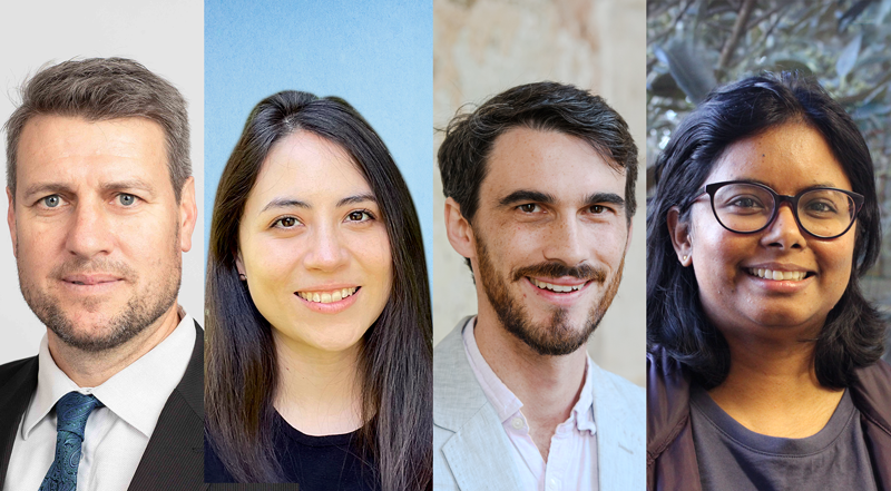
Paul Keall, Emily Hewson, David Waddington and Chandrima Sengupta are among the recipients of the $650 million Medical Research Future Fund (MRFF) National Critical Research Infrastructure initiative. The grant, titled “An AI Platform for Targeted Radiotherapy to Improve Cancer Patient Outcomes”, was publicly announced this week by Minister for Health and Aged Care, The Hon Mark Butler MP.
“These important research projects will help build the infrastructure that will underpin our use of AI in the healthcare system, improving the lives of Australians everywhere.” Said Minister Butler.
April 2024
Trio of Grant Successes
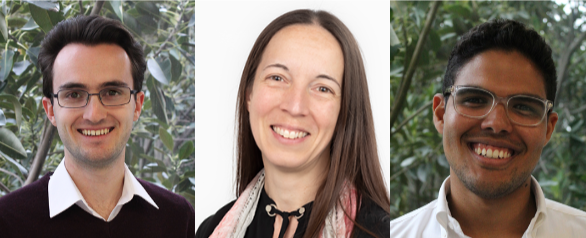
Congratulations to Owen Dillon, Hilary Byrne and Nicholas Hindley (picture L-R), all of whom have been successful in recent funding rounds.
Owen Dillon: Creating the Next Generation in Radiotherapy Imaging with Carbon Nanotube Sources and Photon Counting Detectors. CCNSW Project Grant
Read more: Cancer Council NSW Announcement
Dr Hilary Byrne: How are you breathing today? Embedding functional imaging throughout radiation therapy to improve lung cancer patient outcomes. Cancer Australia ECR PdCCRS grant
Dr Nicholas Hindley: From relativity to respiration: How ideas from Einstein’s general theory enable adaptative radiation therapy for lung cancer patients. Cancer Australia Category A ECR PdCCRS grant
Read More: Cancer Australia Announcement
Jan 2024
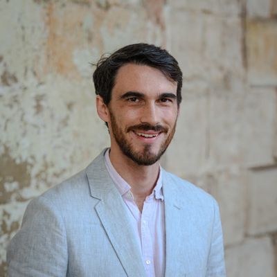
Patent Awarded to David Waddington
Congratulations to David Waddington, who has been awarded a patent for “Systems and methods for susceptibility contrast imaging of nanoparticles at low magnetic fields”. In 2020, David published in Science Advances showing that iron-oxide nanoparticles could revolutionise the contrast possibilities for MRI, especially in low-field, portable setups.
Now, this patent, co-invented with collaborators Matthew Rosen from Massachusetts General Hospital and Zdenka Kuncic from University of Sydney, safeguards the game-changing technique David introduced in that paper.
Given the rapid expansion of the portable MRI market segment, we’re excited for the potential licensing opportunities for this patent. Watch this space!
Read the paper here: https://www.science.org/doi/10.1126/sciadv.abb0998
Nov 2023
EVENT: Research Showcase
You’re invited to join us on Thursday November 9 for an afternoon showcase of the incredible research happening here at Image X. Following the showcase, we are honoured to host a presentation from Professor Laurence Court from the MD Anderson Cancer Centre, University of Texas.
When: Thursday November 9th, from 2:30pm
Where: Seminar Room 422, Level 4, Biomedical Building 1 Central Avenue Eveleigh, NSW 2015
RSVP/More Info: register for free via Eventbrite by Nov 8th: Register + More Info
Sep 2023
Ten for ten at EPSM!
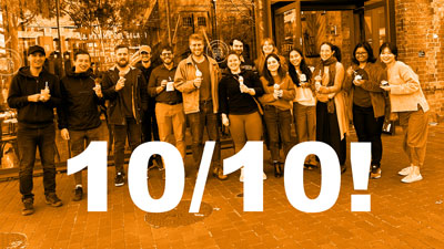
Typically at conferences abstracts are independently reviewed and scored. Those more highly ranked are given oral presentations. Image X are proud that for the upcoming EPSM conference in Christchurch, all 10 of our team’s submitted abstracts all received oral presentations. 10 oral presentations from 10 submitted abstracts is a great result! Pictured left, a whole-institute gelato celebration.
March 2023
Benjamin Lau awarded Fulbright Fellowship
Congratulations to PhD candidate Benjamin Lau, who has been awarded a Fulbright Scholarship. Ben’s research focuses on the development of the next generation of 4D imaging systems for radiotherapy. Mentored by Dr Tess Reynolds, Benjamin’s research aims to reduce radiation exposure and imaging time whilst improving patient outcomes in lung cancer treatment, the leading cause of cancer death and one of the most challenging areas to treat. As a Fulbright Scholar, Benjamin will join the Advanced Imaging Algorithms and Instrumentation Laboratory at Johns Hopkins University, where he will undoubtedly expand his expertise and continue his groundbreaking work.
Read the more here: https://www.sydney.edu.au/news-opinion/news/2023/03/03/2023-fulbright-scholars.html
January 2023
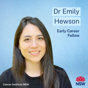
Emily Hewson awarded Cancer Institute NSW Fellowship
Congratulations to Emily, who has been awarded the prestigious fellowship for her project Enabling precise radiotherapy to improve outcomes for advanced lung cancer patients.
“Breathing motion causes radiation to miss the tumour and instead damage healthy tissue, decreasing the rate of cancer curing and the patient’s quality of life. This problem is even more critical for patients with advanced cancer as they require multiple tumours to be treated simultaneously.
I have invented a method to solve the problem of accurately treating cancer patients who have multiple tumours with radiotherapy. I will build upon my adaptive radiotherapy technology to address the complex challenge of respiratory motion.”
Read the more here: https://www.cancer.nsw.gov.au/research-and-data/grants/grants-we-ve-funded/career-support-grants/2023-career-support-funds-granted#Hewson
Oct 2022
Unique partnership revolutionising surgical imaging with robotics
A research collaboration between a world-leading medical device company Siemens and an award-winning physicist Tess Reynolds is promising clear, real-time imaging of moving organs during complex surgeries, de-risking these procedures and greatly improving patient outcomes.
View the full article here: https://www.sydney.edu.au/medicine-health/news-and-events/2022/10/12/unique-partnership-revolutionising-surgical-imaging-with-robotic0.html
Sep 2022
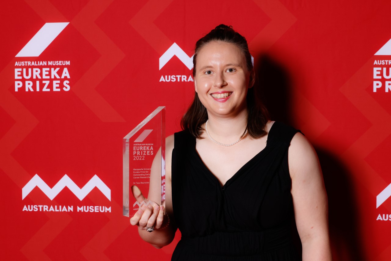
Eureka Prize Winner – Congratulations Dr Tess Reynolds!
Dr Tess Reynolds has won the Eureka Prize for Outstanding Early Career Researcher. Tess received the award for her large body of innovative work. By developing technology to better guide robotic imaging during surgery, Dr Tess Reynolds is improving the view for surgeons as well as outcomes for patients. Partnering with the world’s largest medical device company, her pioneering techniques offer clearer, more complete images for complex cardiac and spinal surgery.
View the full list of winners here: https://australian.museum/get-involved/eureka-prizes/2022-eureka-prize-winners/
July 2022
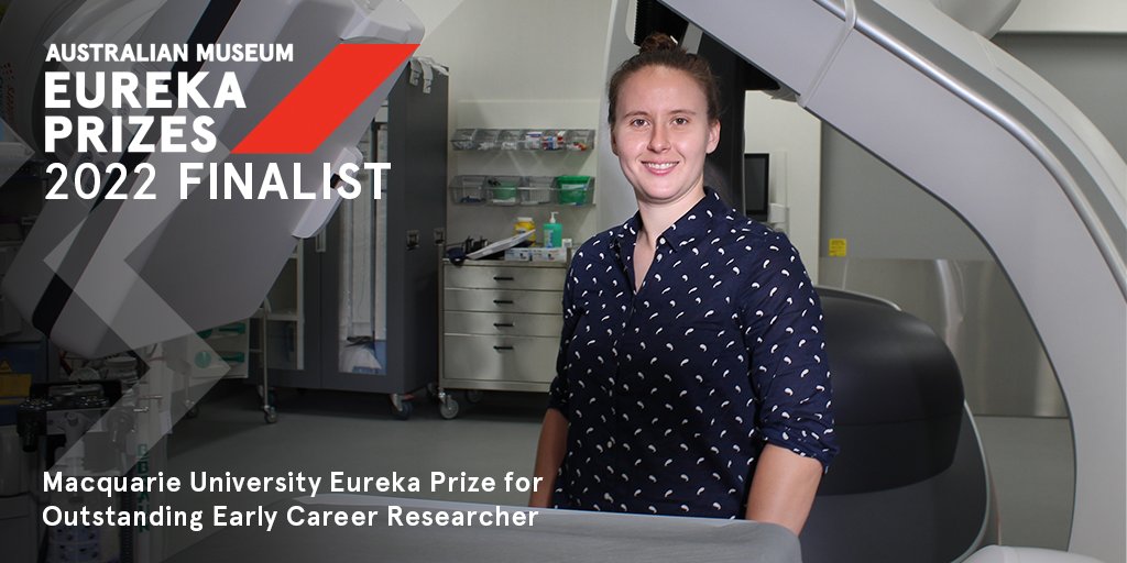
Eureka Prizes Finalist – Congratulations Dr Tess Reynolds
The institute is proud to see Tess nominated as a finalist for Outstanding Early Career Researcher for the second time in as many years.
Dr Tess Reynolds’s ACROBEAT technology enables 3D imaging hardware to calculate heart movement and select the best time to acquire clear images. This has the potential to transform 3D image guidance and change the way we treat cancer and cardiovascular disease.
The technology can be used to guide radiotherapy to organs impacted by cardiac motion and allows surgeons to scan patients mid-operation, to check that heart valves or pacemakers have been placed accurately.
In addition to this, Tess has developed techniques to extend the intra-operative field-of-view, allowing long anatomical sites such as the spine to be captured in a single 3D image during surgery, providing guidance and ensuring accurate hardware and instrumentation placement during complex procedures.
Read the university’s statement here: https://www.sydney.edu.au/news-opinion/news/2022/07/27/2022-eureka-prize-finalists-announced.html
June 2022
James Grover featured in Physics World
Congratulations to James Grover, whose first paper on machine learning for ventilation imaging, was reviewed and highlighted by PhysicsWorld.
James’ work applies a fast-growing computational method, machine learning, to the task of generating functional images from CT scans. For lung cancer patients receiving radiation therapy, this means that the healthier areas of the lung can be identified, and preferentially spared from higher doses of radiation. If the healthier lungs are spared, data suggests that there will be fewer side effects to patients from the radiation therapy treatment.
Read the article here: https://physicsworld.com/a/neural-network-generates-lung-ventilation-images-from-ct-scans/
May 2022
Hilary Byrne awarded REDI Industry Fellowship
Congratulations to Hilary Byrne, who has been awarded an industry fellowship with 4DMedical, to help translate CT Ventilation Imaging for industry.
Dr Hilary Byrne, a Program Manager and Research Fellow at the ACRF Image X Institute in the Faculty of Medicine and Health, will undertake a 12-month project with 4DMedical, the technology innovator delivering breakthrough four-dimensional lung imaging capability, transforming diagnosis and surveillance.
Dr Byrne will focus on resolving clear clinical needs spanning conditions such as COPD, lung cancer, cystic fibrosis and long-COVID, and by expediting regulatory approvals and commissioning of prototype scanning platforms enabling better treatments for patients.
The REDI Fellowship program is part of MTPConnect’s REDI initiative funded by the Australian Government’s Medical Research Future Fund (MRFF). It will bring researchers, clinicians and MTP professionals together for up to twelve months to work on priority medical research projects.
Read more about the REDI Fellowships here: https://www.mtpconnect.org.au/Story?Action=View&Story_id=474
April 2022
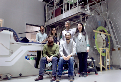
MRI Linac team published in Nature Reviews Clinical Oncology
A rapidly growing body of evidence suggests that MRI-guidance can reduce radiation-treatment-related toxicity by more than 50% as compared to conventional CT guided treatments.
Published last week in leading oncology journal Nature Reviews Clinical Oncology, our researchers have collaborated on an important and timely resource for oncologists. The invited paper Integrated MRI-guided radiation therapy: Opportunities and challenges comes at a time when a new technology, MRI-guided radiation therapy (MRIgRT) is rapidly shifting the field of cancer radiation therapy. Led by Institute Director Professor Paul Keall, this high profile review brought together international research leaders from Europe, the USA and Australia.
“MRI-Linacs are new cancer treatment systems that have integrated an MRI scanner and linear accelerator, are rapidly being clinically deployed: however the opportunity for outcome improvements comes with the challenges of added complexity and cost.” says Professor Keall.
In addition to Prof Keall, five researchers from Image X contributed to the work; Brendan Whelan, Caterina Brighi, David Waddington, Shanshan Shan and Paul Liu.
Read the paper Integrated MRI-guided radiation therapy: Opportunities and challenges for free here: https://rdcu.be/cLF8T
February 2022
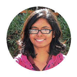
Chandrima Sengupta featured in the University of Sydney’s Clinical Trials Support Office newsletter
Chandrima has an excellent knack for conveying the complexity of her work into accessible terms. Stemming from a presentation she gave last year at a Clinical Trials Support Office event, she was interviewed for their newsletter:
The Australian Cancer Research Foundation (ACRF) Image X Institute is a centre for innovation in radiation therapy and cancer imaging technologies and is part of the Faculty of Medicine and Health. Postdoctoral fellow Dr Chandrima Sengupta shares the story of her journey in medical research thus far and tells us about the exciting research she’s conducting.
Can you tell us about your studies and career thus far?
I was born and brought up in Kolkata, India, and completed my BSc and MSc in physics at the University of Calcutta.
I moved to Australia in 2015 to do a PhD in nuclear physics at the Australian National University. This was a great experience for me to explore the new culture and people around me, learning science and new techniques, and of course learning to speak in English 😊.
I joined the ACRF Image X Institute as a Postdoctoral Fellow in 2019 and since then have been working on improving radiation therapy treatment by developing and implementing new imaging and treatment technologies.
What made you decide to go into medical research?
Medical research is a field where you can directly see the impact of your work. It’s a huge satisfaction to see work that starts as some mathematical equations being used to treat and improve the lives of cancer patients.
With the new proton therapy centre being built in Adelaide, I think from a career perspective there will be lots of research opportunities in this field in Australia. Medical research is being heavily promoted by the Australian Government and the future looks bright.
Can you tell us about the trials you are currently working on and how the outcomes will impact on people’s lives?
I am currently involved in a few clinical trials, but let’s focus on the TROG 17.03 LARK trial (Liver Ablative Radiotherapy utilising Kilovoltage Intrafraction Monitoring).
The human body is dynamic. During radiation therapy treatment, some tumours, such as liver tumours, can move up to a few centimetres from a planned position, mostly due to breathing motion. This is a problem because if we don’t know the precise tumour position during a treatment, the radiation beam may not hit the tumour and may radiate the nearby healthy tissues.
We have a technology developed at the University of Sydney called Kilovoltage Intrafraction Monitoring, or KIM, that can detect the tumour motion and guide the treatment team to correct for the motion by shifting the radiation beam or the treatment couch.
In the LARK trial, our aim is to quantify the tumour tracking accuracy of KIM and delivered dose with KIM guidance, as compared to the standard of care treatment.
We have shown that KIM can be easily integrated with the current standard of care radiation therapy systems, and no extra cost is involved for its clinical implementation. This is significant because existing tumour motion monitoring technologies are way too expensive and hence inaccessible to most cancer patients.
Thus far we have found that with KIM guidance we can deliver radiation to within 1.5 mm accuracy, even if the patient is moving, and the average treatment time is shorter than the current standard of care treatment time.
Our ultimate aim is to deliver a higher dose of radiation with higher accuracy in smaller treatment time at no extra cost, making radiation therapy treatment easier and accessible to all cancer patients and reducing burden on the health system.
What excites you about your role in clinical trials?
We are currently running the LARK trial at three sites across Australia, with three new sites soon to be active. When visiting other sites I get to know the radiation therapy treatment workflow at these centres, as well as the people involved. This in turn gives me ideas for further improvements in treatment workflow and opens up the possibility of new collaborations.
And what are some challenges?
While running a clinical trial is a fulfilling experience, it is also a challenging one with plenty of responsibility – we need to ensure that our technology is safely implemented in the clinic.
I am the first point of contact when any decision needs to be made for KIM operation before and during a patient’s treatment. This was stressful when I first started my job, but having found I’ve been able to handle it over these past two years – with lots of help from my supervisors Professor Paul Keall and Dr Doan Nguyen – I now enjoy facing and solving all these difficult problems.
What’s your top tip for people wanting to get into medical research?
At the end of my PhD, when I was applying for postdoc positions in medical research, I got a few rejections saying that while I had a great profile, I didn’t have relevant experience in medical physics. So, if you are from a different research field like me, it may be hard at the beginning to get a position in medical research, but don’t give up.
As mentioned earlier, there are lots of interesting topics in this field and we will see many more in the near future. Dive into it!
Lastly, here’s a list of clinical trials, researchers from Image X are conducting:image-x.sydney.edu.au/clinical-trials/
See the full newsletter here: https://sway.office.com/TzwgbxrNHCLqtDpj?ref=Link
December 2021
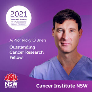
Professor Ricky O’Brien Recognized in the NSW Premiere’s Research Awards
Professor Ricky O’Brien has been annoucned the winner of the Outstanding Cancer Research Fellow – Career Development Fellow in the NSW Premier’s Research Awards. This fellowship award acknowledges Professor O’Brien’s outstanding work in progressing imaging technologies for cancer.
Professor O’Brien’s fellowship is directly benefiting people with cancer, by delivering shorter scan times and better image quality. Together these can contribute to more accurate results and faster treatments. Changes to scans, such as lower pre-treatment imaging doses, also have the potential to make scans safer for patients.
Read the announcement here
Sep 2021
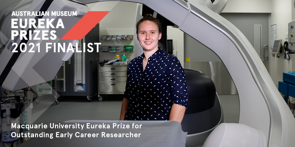
Eureka Prize Nomination – Dr Tess Reynolds
Tess Reynolds has been named a top-3 finalist for the 2021 Australian Museum Eureka Prize for Outstanding Early Career Researcher. The Eureka Prizes are Australia’s most comprehensive national science awards, honouring excellence in research and innovation, leadership, science engagement, and school science. Tess was nominated by the University of Sydney for her work developing ACROBEAT (Adaptive CaRdiac cOne BEAm computed Tomography). ACROBEAT is a new technology that enables imaging and treatment hardware to beat in sync with the patient, delivering clearer, faster, and safer medical images. ACROBEAT has the potential to transform 3D image guidance and change the way we treat cancer and cardiovascular disease. The award winners will be announced on October 7th
Read the University of Sydney announcement here.
May 2021
Funding Success for Dr Doan (Trang) Nguyen
Congratulations to Dr Nguyen, who will use artificial intelligence to enhance radiotherapy effectiveness, thanks to $449,316 of support from Cancer Council NSW.
Harnessing the power of artificial intelligence, in this project Dr Doan Trang Nguyen and her team will implement “beam’s-eye-view” tracking technology during radiation treatment for prostate, liver and pancreas cancer patients. With beam’s-eye-view tracking, the cancer is tracked at all time during radiation therapy treatment, ensuring high accuracy and high precision treatment. For the patients, this means their cancer is hit and destroyed while their healthy tissue and organs are protected from damage.
Read more here.

May 2021
Image X Researchers Awarded by AAPM
Congratulations to two of our researchers, who have been recognized for their innovations in physics in medicine. Emily Hewson and Dr Tess Reynolds have each been given top awards by the American Association for Physics in Medicine (AAPM), and as a result their research will be under the spotlight at the world’s largest medical physics conference this winter.
Dr Tess Reynolds has won the Jack Fowler Early-Career Investigator Competition for her work to improve the way we can image the spine during surgery. To cross-check the accuracy and success of certain spinal procedures, a 2D image of a small section of the spine is taken during surgery.
Tess has developed a way to take an image that covers up to 5x more of the length of the spine, in 3D, to supplement the standard 2D images. This novel imaging modality could drastically reduce the necessity for repeat procedures, which are commonly needed in spinal surgery. The work arose from an academic-industry partnership between the University of Sydney, Johns Hopkins University and Siemens Healthcare.
“The academic side of the collaboration with Johns Hopkins was formed during the height of the pandemic last year, so its really exciting to see how far this work has been able to come and to be recognised by the AAPM in such a short and strange period of time.”, says Tess.
PhD Candidate Emily Hewson received the Best in Physics award for her abstract titled Real-Time Dose-Optimized Multi-Target MLC Tracking for Locally Advanced Prostate Cancer. This work was funded by Cancer Council NSW. Emily’s world-unique software brings us closer towards treating multiple tumours simultaneously with radiation. This technology would improve Radiation Therapy for hundreds of thousands of patients with locally advanced prostate, lung or liver cancer.
Tess and Emily’s submissions were judged in the top 1% of all submissions, an outstanding achievement.
“Each year the Image X Institute has a strong presence at the conference, but to see two incredible young women receiving such high accolades early in their academic careers makes 2021 that much more exciting.” says Professor Paul Keall, Director of the ACRF Image X Institute.
March 2021
ADAPT Trial – Early Results
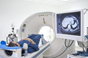
Early results of the ADAPT clinical trial show that faster imaging with a lower dose + higher quality is possible, bringing improved care to future lung cancer patients. In this trial, Respiratory Motion Guided (RMG) 4DCBCT will be implemented for the first time on lung cancer patients. RMG-4DCBCT adapts the image acquisition as the patient’s breathing changes (i.e. if the patient breathes faster, imaging data is acquired faster). By adapting the acquisition to the dynamic patient we are able to acquire personalised images of a patients lungs for radiotherapy treatments.
Read the article here.
Oct 2020
Emily Hewson featured in Physics World
PhD Candidate Emily Hewson has been interviewed by new website Physics World about her recently published article assessing multileaf collimator tracking and gating of the radiation therapy beam during treatment.
“Our implementation of KIM to monitor tumour motion, combined with either gating or MLC tracking improves the availability of intrafraction motion adaption for all clinics with standard treatment machines,” says Hewson. “One of the major barriers to implementing real-time adaptive radiotherapy in many countries has been a lack of finances and resources. The adaptive methods we compared could potentially overcome these obstacles and bring intrafraction motion adaptation into standard clinical practice at any cancer treatment facility that treat patients using a modern linear accelerator.”
Read the article here.
Dr Paul Liu featured on Phyics World
Paul Liu, has been featured on the prominent Physics news website for his work: MRI-Linac enables simultaneous MLC tracking of two moving targets
Paul has described the use of an MRI-Linac to simultaneously track the motion of two treatment targets. “There are many radiotherapy cases that involve simultaneous treatment of multiple targets,” he said, citing examples such as locally advanced prostate or lung tumours, and oligometastases.
Read the article here.
July 2020
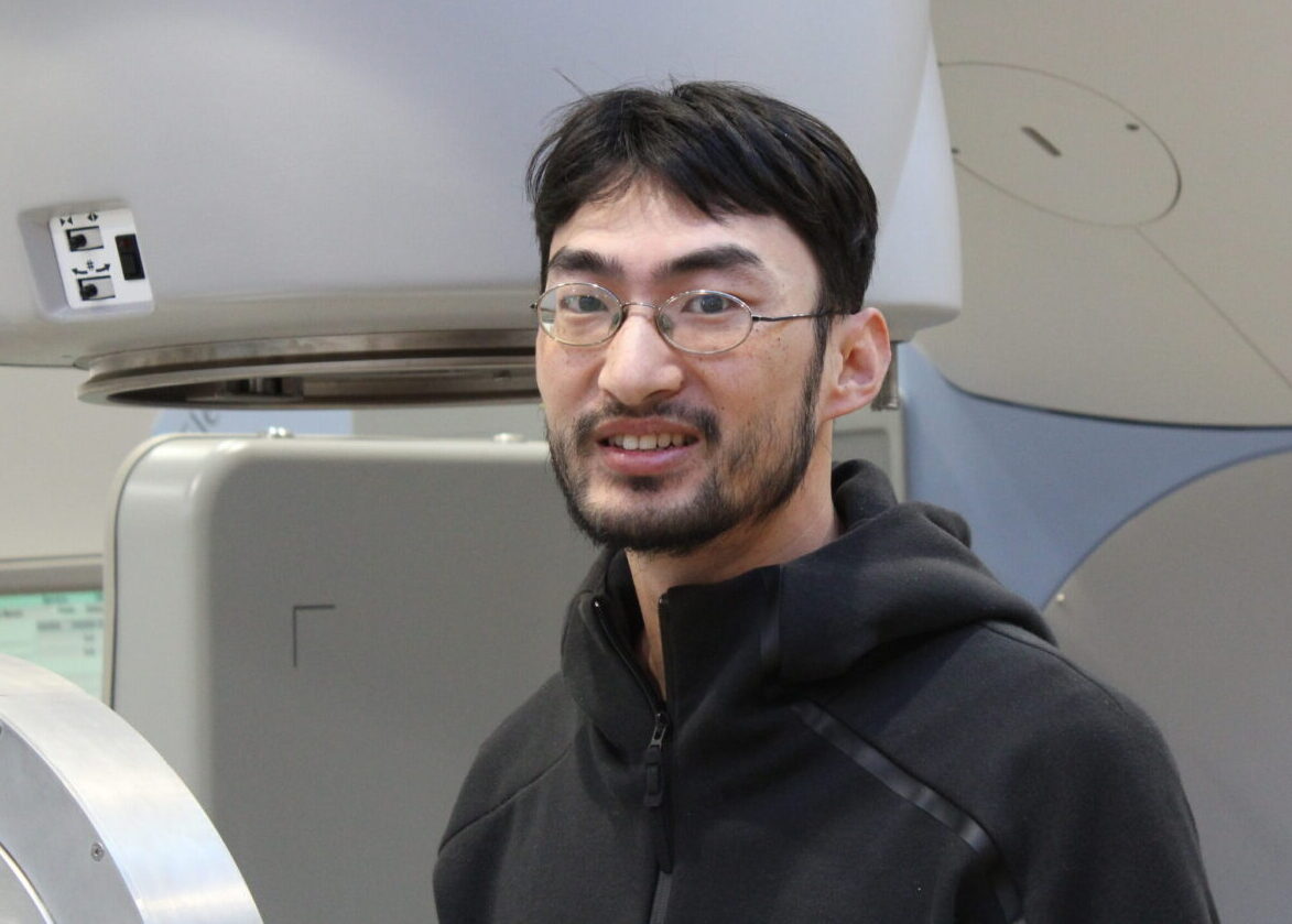
Study identifies missing piece needed for lower-cost, high-quality MRI
David Waddington‘s paper “High-sensitivity in vivo contrast for ultra-low field magnetic resonance imaging using superparamagnetic iron oxide nanoparticles” has been published in Science Advances. The research was completed during David’s Fulbright scholarship, in collaboration with friend of the institute, Zdenka Kuncic (University of Sydney) and Matt Rosen (Massachussetts General Hospital), amongst others. The paper has been featured in the university’s News and Opinion here, and Medical Xpress here.
July 2020
$2.1M awarded by NHMRC.
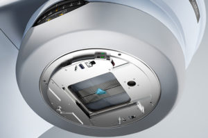 Professor Paul Keall has been awarded a $2.1M investigator grant for the project Cancer Imaging and Targeted Radiation Therapy: From Discovery to Clinical Practice. This funding will allow us to advance real-time targeted radiotherapy by better imaging, targeting and adapting radiation therapy to moving tumours, and sparing surrounding healthy organs. It will also enable us to explore the delivery of personalised anatomically and physiologically targeted radiation therapy by tailoring treatments to individual patients.
April 2020
Record Number of Early Career Fellowships to improve Cancer Radiation Therapy with MRI.
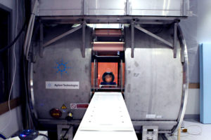 A trio of early career research fellows from the ACRF Image X Institute at the University of Sydney will begin cutting-edge research missions to improve cancer radiation therapy this year, thanks to support from Cancer Institute NSW and the NHMRC. Dr Brendan Whelan, Dr David Waddington and Dr Paul Liu will each be tackling unique challenges associated with the Australian MRI Linac, a powerful experimental cancer radiation therapy system based at Liverpool Hospital.
Read the press release here.
Learn about The Australian MRI Linac Program here.
18 December 2019
Remove the Mask receives $600,000 funding from Cancer Australia
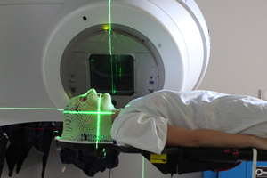
The Morrison Government has today announced the awarding of $8.9 million for cancer research in Australia, through Cancer Australia’s Priority-driven Collaborative Cancer Research Scheme (PdCCRS).
Remove the Mask received $600,000 as part of the funding round, which will be used to develop a surface guidance technology for the treatment of head and neck cancers that will reduce anxiety and stress in radiotherapy patients, by Professor Paul Keall at the University of Sydney.
Read the press release here.
Learn about Remove the Mask here.
Dec 2019
Prize winning PhD Candidates at MedPhys 2019
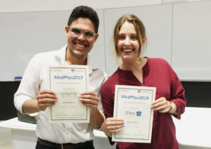 It is with great pride that we congratulate our higher degree students for their exceptional presentations at Medphys 2019, the annual event hosted by The Australasian College of Physical Scientists & Engineers in Medicine (ACPSEM) NSW Branch.
Natasha Morton was awarded Best Postgraduate PhD for Creating clearer images for clinical use: syncing patient breathing to CT imaging.
Nicholas Hindley was awarded Most Outstanding Presentation for Real-time direct diaphragm tracking during lung cancer radiotherapy.
October 2019
Congratulations Nicholas Hindley – Fulbright Scholarship Awarded
Nick Hindley has received the prestigious Fulbright Foreign Student Program.
He will spend part of next year at Massachusetts General Hospital/Harvard Medical School, in addition to visiting other sites where machine learning and image reconstruction are strong themes.
September 2019
Respiration-guided imaging method could set a new benchmark for cancer imaging.
 Researchers at the University of Sydney have used a cancer patient’s breathing signal to guide the acquisition of a pre-treatment CBCT scan, commonly used when planning radiotherapy for cancer patients. This milestone scan marks the beginning of the ADAPT clinical trial, which is expected to demonstrate a dramatic improvement in image quality whilst reducing imaging time and radiation dose by 75% for cancer patients.
In current cancer imaging, patients are imaged continuously as they breathe. The motion of the lungs during imaging results in blurring and streaks which degrade the clarity of the image. In the ADAPT Trial, the patient’s breathing signal is used to adapt and move the imaging machine and also to determine the optimum time in the breathing cycle to acquire the image, resulting in dramatically clearer and more accurate images, with a lower radiation dose delivered to the patient.
“Clearer images are integral to providing even more effective radiotherapy treatment. The better we can see the cancer, the more accurately we can treat it.” says A/Prof Ricky O’Brien, who is leading the trial at the University of Sydney in collaboration with Liverpool Hospital. The technology was the subject of an NHMRC grant and achievement award in 2011 which topped 3500 grant applications.
“The ADAPT Trial is the touchpoint where the core concept of the ACRF Image X Institute’s Patient Connected Imaging Program begins to have a real-world impact on the lives of cancer patients.” Connecting the patient’s physiological signals to the imaging system signifies a major step forward in cancer imaging technology, which can in turn result in a more personalised, effective treatment for each patient.”, says Professor Paul Keall, director of the ACRF Image X Institute at the University of Sydney
The ADAPT trial (full name Adaptive CT acquisition for personalised thoracic imaging: A Phase 1 Pilot Study on the Use of Respiratory Motion Guided 4DCBCT for Lung Cancer Radiotherapy) is the result of collaborations forged between ACRF Image X Institute, the Netherlands Cancer Institute and Liverpool Hospital.
April 2019
Crowdfunding – Remove the Mask
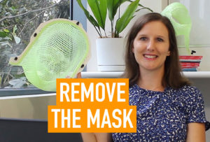 We’re joining forces with cancer survivors to crowdfund a project which is set to revolutionise radiation therapy for people with head & neck cancer. If successful, we’ll be on the path to reducing the debilitating anxiety and fear experienced by hundreds of thousands of people every year who must wear a mask for their radiation therapy.
Through our work on other cancer sites including breast, prostate, liver and lung, we’ve already developed two of the technologies needed to be able to remove the mask. With your help, we can develop the final piece of the puzzle – surface mapping technology, which will monitor the position of the patient.
You can make a donation or keep up to date with the campaign here.
14 April 2019
Physics World – Feature Article
Our work to improve cardiac diagnostic imaging is in the spotlight, with a feature article on our recent ACROBEAT publication.
“Cardiac images reconstructed using the conventional protocol contained streak and blurring artefacts for all three ECG traces. In all ACROBEAT simulations, these artefacts almost completely disappeared.”
Read the full article here.


