South Eveleigh Biomedical Building
Our headquarters, located in the vibrant South Eveleigh precinct are a stone’s throw from Redfern station. Our space comprises closed plan offices with multiple breakout spaces to foster both concentration and collaboration. Our headquarters are also home to:
- Robust computing resources
- Versatile laboratory/workshop space
- Wet lab access
- Hybrid seminar and workshop facilities
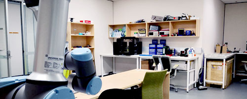
Workshop/Lab
- 3D printing lab (see printers below)
- Robotic arm (UR16e UR3e)
- Drill press
- Soldering station
- Hand tools
- Power tools
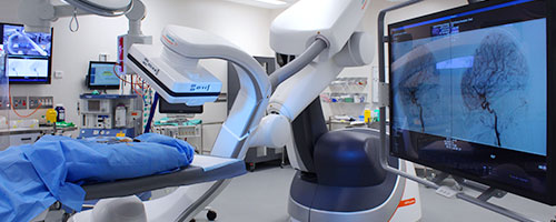
Hybrid Theatre, Charles Perkins Centre
The Hybrid Theatre has a vast array of research and training capabilities across a range of medical disciplines, such as interventional cardiology, neurosurgery, cardiothoracic surgery, laparoscopic surgery, cancer treatment planning, and any field where visualizing devices or contrast is paramount.
The first instrument of its type in Australia, the Artis Pheno offers exceptional CBCT imaging quality in a wide range of clinical cases. The robot-supported angiography system was developed for use in minimally invasive surgery, interventional radiology and interventional cardiology.
Additional equipment
- Siemens SC2000 ACUSON
- Image intensifier
- IT monitoring equipment
- Anaesthesia machine
- Heart-lung machine
- Robotic surgical systems
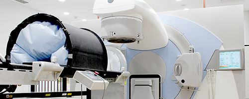
Nelune Comprehensive Cancer Centre
Image X has secured access to a dedicated research bunker at Nelune Comprehensive Cancer Centre. This dedicated suite, designed to mirror its neighbouring treatment bunkers, is specifically tailored for radiation therapy research,
- Elekta Linear Accelerator with integrated cone beam CT imaging capable of producing capable of producing high energy x-ray and electron beams.
- Photon Counting Detector (PCD) which is able to see “in colour” across the x-ray spectrum, giving better contrast between different types of tissue.
- Carbon Nanotube (CNT) source which can be digitally focussed to acquire 3D x-ray images far faster than mechanically moving conventional sources.
- Wireless x-ray detector, which we can move to various positions to simulate acquiring x-ray images with novel system geometries.
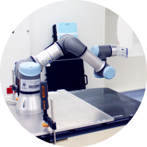
Robotic systems
- Our six degree-of-freedom robotic motion system for quality assurance of real-time image-guided radiotherapy consists of:
- 2 commercially available robotic arms,
- an acrylic phantom with embedded gold fiducial markers,
- a custom base plate for secure mounting to treatment systems,
- Image X developed control software that reproduces measured tumour motion traces.
Phantoms
To facilitate our research, we use a range of off-the-shelf and custom-phantoms. Some notable examples include:
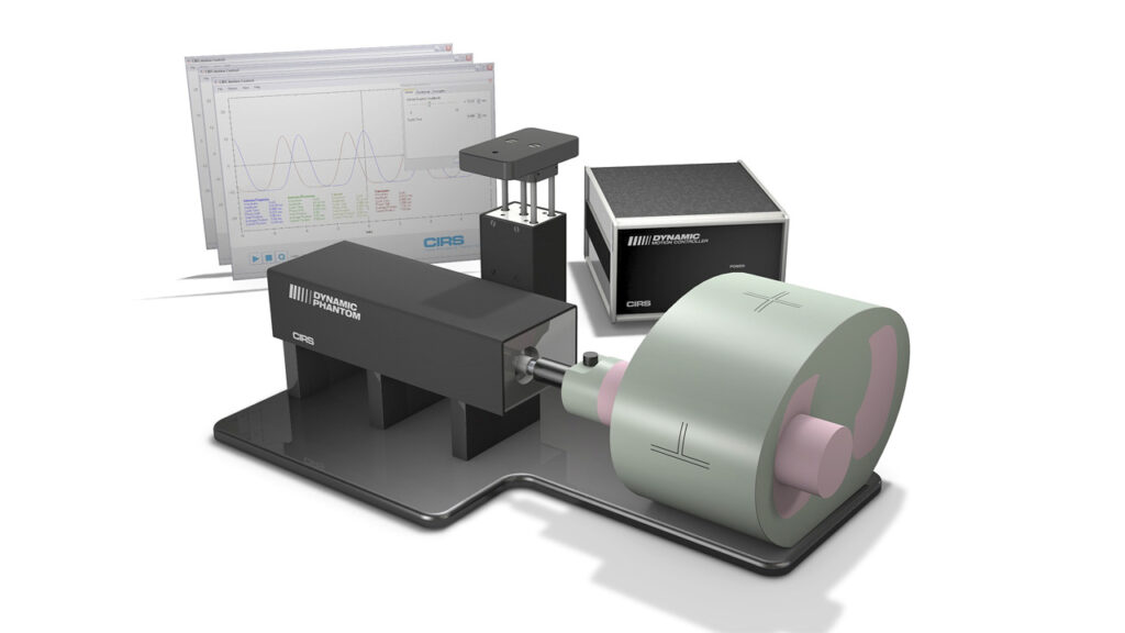
The CIRS Dynamic Cardiac Phantom simulates the realistic motion of an average human heart. It provides known, accurate and repeatable 3D motion of a solid heart model inside the tissue-equivalent thorax phantom.
The CIRS Dynamic Thorax Phantom provides known, accurate and repeatable 3D target motion inside a tissue equivalent phantom for investigating and mnimising the impact of tumour motion in the lung.
T-Rex is a life-size 3D-printed phantom based on a clinical CT scan of the thorax from a patient with lung cancer. It was assembled from bony structures printed in gypsum, lung structures consisting of airways, blood vessels >1 mm, and outer lung surface, three lung tumors printed in nylon, and soft tissues represented by silicone.
The XCAT tool is a CT simulator based on the 4D extended cardiac-torso (XCAT) phantom, a whole-body computer model of human anatomy and physiology. The XCAT software is currently available for download from the Image-X GitHub and is on two Image-X workstations: Costa (Windows) and Goliath (Linux).
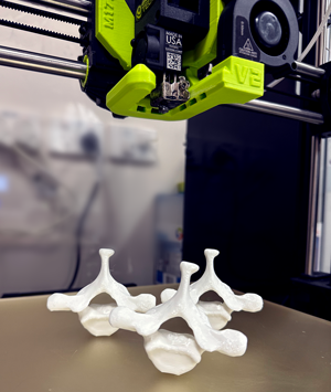
3D printers
The Spineprint Lab at the South Eveleigh Biomedical Building houses a collection of 3D Printers used across different projects:
Stereolithography – FormLabs Form 2
Masked Stereolithography – Phrozen Sonic Mini Resin 3D-Printer
Fused Deposition Modelling – LulzBot TAZ Workhorse