
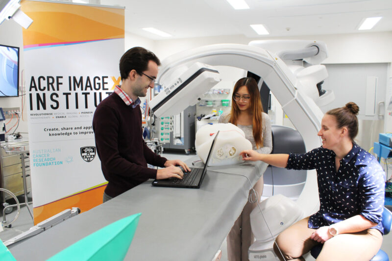
Each year Image X Institute offers a Summer Research Program, running across January and February.
These scholarships provide an opportunity for students studying at the University of Sydney to undertake a summer vacation research project with investigators in our research program.
The projects are especially suited to students with a background in subjects including (but not limited) to:
- Physics
- Mathematics
- Computer science
- Biomedical engineering
- Electrical engineering
Image X prides itself on offering a summer research program filled with opportunities. Students can work onsite amongst our multidisciplinary team, learning about the different projects and careers within our group. We host social activities, tours of our research facilities, as well as an introductory seminar series about radiation therapy and cancer imaging, presented by our star researchers. Many summer students continue working with Image X after their projects, from taking on part time research positions through to commencing PhD projects.
Successful applicants will be invited to choose from the projects below (please note, more projects will be added in the next month). Scholarships will be awarded based on the student’s academic transcript, relevant academic background or any research experience.
Apply for 2027
Applications for scholarships in the 2026/27 summer vacation are open!
Submit your application by September 20th 2026.
Watch a quick video to meet some former summer students here:
Projects in 2027
More projects are being added across September/October
AI-Driven Liver Deformation Tracking for Precision GTV Localisation
Supervisors: Dr. Zhuang Xiong, Dr Chandrima Sengupta & Gregory Willson
Project description:
This project will develop a method to estimate breath-hold liver deformation by registering real-time 2D X-ray images (with the potential to extend to 3D volumetric imaging) to the liver in a 3D abdominal planning CT. Biomechanical modelling will be used to generate realistic liver deformations and constrain the estimated motion to physically plausible patterns. The goal is to track liver deformation and infer the Gross Tumor Volume (GTV) position based on this deformation during radiotherapy using deep learning.
Experience with Python and Python-based deep learning frameworks such as PyTorch is essential, while familiarity with MATLAB is a bonus. We are looking for a self-motivated student interested in medical imaging and deep learning.
Developing deep learning methods for creating rapid radiation therapy treatment plans
Supervisors: Dr Mark Gardner
Project description:
The creation of accurate radiation therapy treatment plans is essential to ensure safe and effective radiation therapy treatments. However creating accurate treatment plans is a complex physics-based optimisation process that can take several hours to complete. As the radiation therapy treatment process as a whole is getting faster with more automation and fewer treatment sessions, the creation of treatment plans is a bottleneck to reducing the time between planning and treatment sessions.
Deep learning methods have been proposed for rapidly creating treatment plans, however the many of the created treatment plans had unacceptably high dose to nearby critical organs. In this project you will investigate novel methods for creating accurate treatment plans using physics and geometry informed deep learning methods.
Experience in python coding and deep learning is preferred.
Deep feature representation learning across DRR and X-ray domains for radiotherapy motion tracking
Supervisors: Dr Chuhan Wang & Dr Chandrima Sengupta
Project description:
Accurate tracking of moving anatomy during radiotherapy is important for improving treatment precision and reducing unnecessary radiation exposure to healthy tissues. However, anatomical motion and differences between digitally reconstructed radiographs (DRRs) and real X-ray images can make reliable tracking challenging.
This project will investigate deep learning methods for learning robust image representations and feature matching across DRR and X-ray images, including variations in image domain and scale. The project will also explore visualization techniques to better understand what image features the model learns, how information is represented across different imaging modalities, and how these representations contribute to anatomical matching and motion-tracking decisions.
The outcomes of this project will contribute to the development of more accurate and reliable real-time motion tracking methods for image-guided radiotherapy, while providing greater insight into how deep learning models interpret radiotherapy images.
Experience with Python, PyTorch and basic deep learning is preferred.
Parahydrogen hyperpolarized nuclei for MRI-guided radiotherapy
Supervisors: Dr Thomas Boele, Dr David Waddington
Project description:
Better diagnostic imaging and biologically targeted radiotherapy can improve cancer patient outcomes. Magnetic resonance imaging (MRI) provides high resolution images with excellent soft tissue contrast for clinical diagnosis. MRI-guided radiotherapy has emerged as a clinical tool in the last decade as treatment devices combining imaging and targeting have become available to oncology departments.
However, MRI is limited by its low sensitivity, inherent to the technique’s reliance on the small net magnetic moment of ensembles of nuclear spins in biological systems. Hyperpolarization techniques can boost the available MR signal by greater than four orders of magnitude to enable imaging modalities that are impractical or impossible with traditional methods.
In this project we aim to build hardware to develop a flexible and low-cost approach based on the transfer of quantum spin order from parahydrogen to the nuclei of interest to enhance access to cutting edge hyperpolarization technology. There are multiple directions of research available for a student interested in exploring the intersection between engineering, quantum physics and medical imaging.
Improved Speech Imaging with Real-Time MRI
Supervisors: Dr David Waddington
Project description:
Speech is a crucial aspect of human communication, yet there is still much to learn about how people shape their vocal tract during speech production. While MRI technology has the potential to provide detailed images of speech, current real-time MRI techniques are too slow to capture the rapid movements involved.
This project aims to utilize advanced reconstruction techniques to enhance the quality and speed of videos showing people speaking while in an MRI scanner. Students with a background in mathematics, engineering, physics, and coding (Python and/or MATLAB) will be well-suited for this project. By the end of the project, the student will produce a comparative analysis of various image reconstruction methods and a set of real-time videos showing human speech. This project is a collaboration with the Discipline of Speech Pathology and the School of Electrical and Computer Engineering.
The impact of low dose CT on CT ventilation image quality in the context of lung cancer screening programs
Supervisors: Prof Paul Keall, Jeremy Lim
Project description:
Computed tomography ventilation imaging (CTVI) is a fast growing clinical modality. CTVI has been applied across respiratory diseases, including lung cancer, interstitial lung disease and idiopathic pulmonary fibrosis. The first CTVI product was FDA-approved in 2023.
Also, increasing are national lung cancer screening programs. The US National Lung Screening Trial found from over 50,000 persons at high risk for lung cancer that screening reduced mortality. Based on the findings, the US National Comprehensive Cancer Network (NCCN) guidelines recommended low dose CT for patients at high risk of lung cancer.
A feature of lung cancer screening programs is that, to balance the costs and benefits of CT imaging, low dose CT scans are used. These scans have increased noise compared with standard CT scans and may impact the quality and accuracy of the CTVI derived from the scans. However, to date, no dedicated studies investigating the impact of low dose CT on CTVI quality have been performed.
Therefore, to address the knowledge gap, the aim of this project is to quantify the impact of low dose CT on CTVI quality in the context of pairing CTVI with lung cancer screening programs.
Towards Reliable Electrocardiographic Imaging
Supervisors: Dr Joana Ooi, Dr Chandrima Sengupta
Project description:
Electrocardiographic Imaging (ECGI) is a non-invasive technique that reconstructs the electrical activity of the heart from body-surface electrocardiogram (ECG) recordings. Cardiac arrhythmias are disorders of the heart’s electrical system that cause abnormal heart rhythms and may require treatment with catheter ablation. Currently, detailed cardiac electrical mapping is typically obtained through invasive electrophysiology procedures, in which catheters are inserted into the heart to identify arrhythmia mechanisms and guide ablation therapy. ECGI seeks to provide similar electrical information non-invasively, with the potential to improve arrhythmia diagnosis, treatment planning, and procedural guidance. However, validation of ECGI remains challenging due to the limited availability of suitable reference data.
This project aims to establish a virtual validation framework for ECGI by generating realistic body-surface ECG data from patient-specific anatomical models with known underlying cardiac electrical activity. The framework will support the development and evaluation of ECGI methods and enable investigation of factors affecting ECGI performance, including modelling assumptions and physiological motion.
Desirable skills: Basic programming experience (Python preferred). Familiarity with signal processing would be advantageous.