Cardiac Radioablation Program
Reducing the impact of radiation on the heart for cancer and cardiac disease patients
The Cardiac Radioablation program is funded by an NHMRC Synergy Grant.
There is currently an opportunity to join the team: https://usyd.wd3.myworkdayjobs.com/en-US/USYD_EXTERNAL_CAREER_SITE/details/Research-Fellow_0109390-1
Background
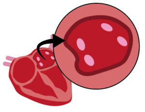
Ventricular tachycardia (VT) and atrial fibrillation (AT) are cardiac conditions that affect over 33.5 million people globally every year. Treatment typically involves invasive heart surgery to manually create scar tissue in specific areas of the heart, to block the abnormal heart signals. Radiation therapy is emerging as a promising new non-invasive treatment option.
Cardiac radioablation is a revolutionary non-invasive method of producing cardiac scarring. The potential of radioablation has been demonstrated through recent clinical trials, however there is considerable room to improve this emerging treatment. To ensure cardiac radioablation treatment efficacy and safety, it is vital that radiation is carefully delivered to the cardiac target, but steered away from the healthy organs and cardiac substructures that are often in very close proximity. A considerable challenge is that the heart is moving with both cardiac contraction and respiration. We are addressing this motion management problem for cardiac radioablation by inventing imaging technologies for the entire treatment pathway from CT simulation, to pre-treatment patient-connected imaging, and during treatment delivery.
Team
| Investigator | Research Institution |
|---|---|
| Prof Ricky O'Brien | RMIT University |
| Prof Paul Keall | Image X Institute - University of Sydney |
| Dr Doan Trang Nguyen | Image X Institute, University of Sydney |
| Assoc Prof Shankar Siva | Peter MacCallum Cancer Centre |
| Dr Tess Reynolds | Image X Institute, University of Sydney |
| Assoc Prof Saurabh Kumar | Western Sydney Local Health District |
| Assoc Prof Lois Holloway | University of New South Wales |
Through a wide array of projects, we’re collaborating with experts across multiple research institutes, clinical sites, and imaging vendors to unleash the full potential of cardiac radioablation:
Accurate CT imaging in the presence of respiratory and cardiac motion (Keall, O’Brien)
In a successful collaboration with Siemens, we’ve countered motion artefacts in images, caused by the patients breathing. Building on this, we’re integrating cardiac motion by adjusting CT table motion and image capture based on real-time respiratory and cardiac signals. This technology can also predict errors from respiratory and cardiac data, pausing the CT beam during anomalies for accurate imaging.
Using an MRI-Linac for real time cardiac tracking and substructure avoidance (Holloway, Keall)
This project expands on our technology to track and monitor the motion of the ablation target in real-time during treatment. The work takes advantage of the exquisite image quality made available by MRI scanners. This research is being conducted in collaboration with the Australian MRI Linac Program, on a novel MRI-Linac (one of only 4 research systems in the world) located at Liverpool Hospital.
Cardiac and respiratory imaging on the treatment table (O’Brien, Reynolds)
Utilising 4DCBCT technology, in this project patients will be positioned for treatment with time-resolved CT images, captured during breathing. Sequential display of these images aids treatment alignment to the planned area, and shows clinicians when re-adjusting is needed. Validation will involve cardiac phantom tests to assess imaging precision.
Understanding the dose response to cardiac substructures in our database of >30K breast and lung cancer patient treatments. (Holloway, Nguyen)
This work utilises patient treatment and follow-up data to assess heart and lung toxicity, quantifying toxicity tolerance. Leveraging the AusCAT network’s data, encompassing 30,000 breast cancer patients’ radiotherapy data, we’ll combine breast and central lung cancer patient data, enhancing it with computer-generated heart contour algorithms. The algorithms will segment heart components, from chambers to arteries, improving accuracy for smaller structures. By analysing treatment plans and patient follow-up, the study will establish dose-response relationships for different heart structures, determining damage probability thresholds.
Integrating EP mapping with treatment planning for more accurate treatments (O’Brien, Reynolds)
EP mapping is used in cardiology to identify the target to ablate. This needs to be combined with the 4D images used to treat patients in radiotherapy. We’re researching into fusing CT cardiac imaging with catheter EP mapping for treatment planning and treatment delivery.
Treatment delivery with cardiac substructure tracking (Keall, Nguyen)
We have already pioneered technology to track the motion of tumours for prostate, lung and liver cancer. Our next goal is to provide, for the first time, information to the radiation therapist on the motion of the ablation target during treatment. This will allow therapists to stop treatment and reposition the patient if the targets move too far away from the treatment beam. We’re working to extend these technologies to include the real-time tracking of the cardiac substructures for cardiac radioablation, and for cancer treatment as well.
Animal studies to determine optimal dose response (Kumar, Siva)
This is a crucial quantitative dose finding animal study to determine the dose required to cause an electrical block or reprogramming for ventricular and atrial targets in the heart. In previous animal studies conducted in Europe, it was assumed that scarring was the mechanism for treatment of AF/VT while in these studies we will be looking for evidence of electrical circuit reprogramming as well as scarring. In our experiments, a diseased model for VT/AF will be developed and verified at the Westmead Cardiology Animal Catheter Lab which has previous expertise with these models as well as animal and cardiac facilities to perform this study
Pioneering human treatments in the Australian healthcare system (Kumar, Siva)
This project aims to expand cardiac radioablation accessibility nationally. In 2020, Dr Shankar Siva used cardiac radioablation to treat the first Australian patients under compassionate grounds. Treatment complexity demands collaboration between cardiology and radiation oncology, which hinders less-equipped centres. The project aims to streamline protocols and knowledge transfer, merging technologies and discoveries, to facilitate cardiac radioablation implementation across multiple Australian centres, addressing treatment challenges.
Contact
For more information about the program and projects, contact Professor Ricky O’Brien: ricky.obrien@rmit.edu.au
If you’re seeking a new challenge, Image X welcomes expressions of interest from researchers with expertise in fields including algorithm development, machine learning, advanced hardware use, and clinical translation across all imaging modalities are welcomed. Email image-x.contact@sydney.edu.au.