Image X Interventional Imaging is an international academic-industry-healthcare program led by Dr Tess Reynolds. It is dedicated to delivering innovative robotic imaging and novel 3D-printed solutions to aid in the diagnosis and treatment of disease spanning cardiology, neurology, oncology, radiology, and orthopaedics.
Project highlights
Extending the intraoperative field-of-view
Intraoperative 3D Cone Beam CT (CBCT) imaging has revolutionised interventional procedures. However, a critical barrier to continued expansion of the technology is the small field-of-view, currently preventing long anatomical sites from being captured in a single 3D image intraoperatively.
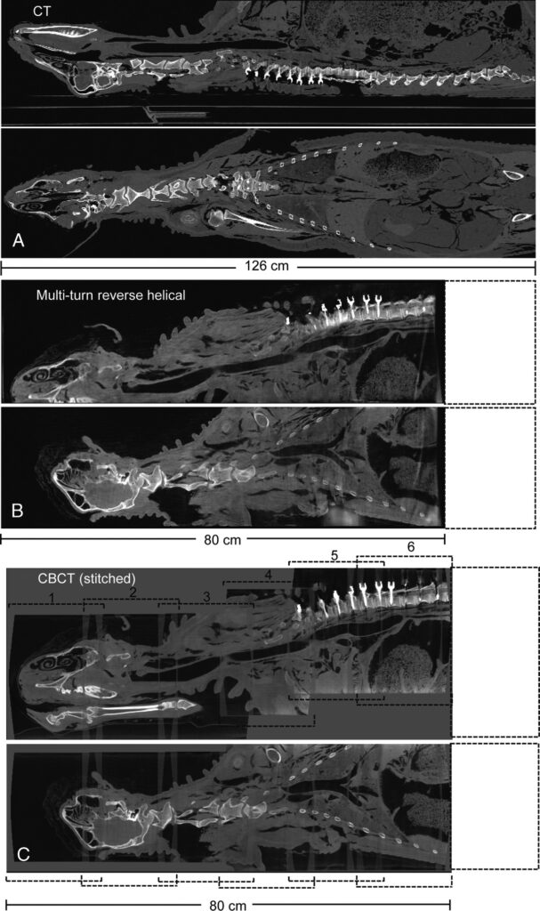
Working in collaboration with Siemens Healthineers and Johns Hopkins University, Image X Interventional Imaging has developed and implemented a new CBCT imaging technique that increases the intraoperative volumetric field-of-view to over 1m. The imaging trajectory is known as a multi-turn reverse helical (demonstrated in the video below Insert video of image acquisition – multi-turn reverse helical), and combines continuous gantry rotation with table translation to bring CT-like longitudinal coverage into the operating room.
An example of multi-turn reverse helical images from a sheep cadaver undergoing a pedicle screw fixation are shown below. (from: https://journals.lww.com/investigativeradiology/Fulltext/9900/Extended_Intraoperative_Longitudinal_3_Dimensional.15.aspx

3D Printing
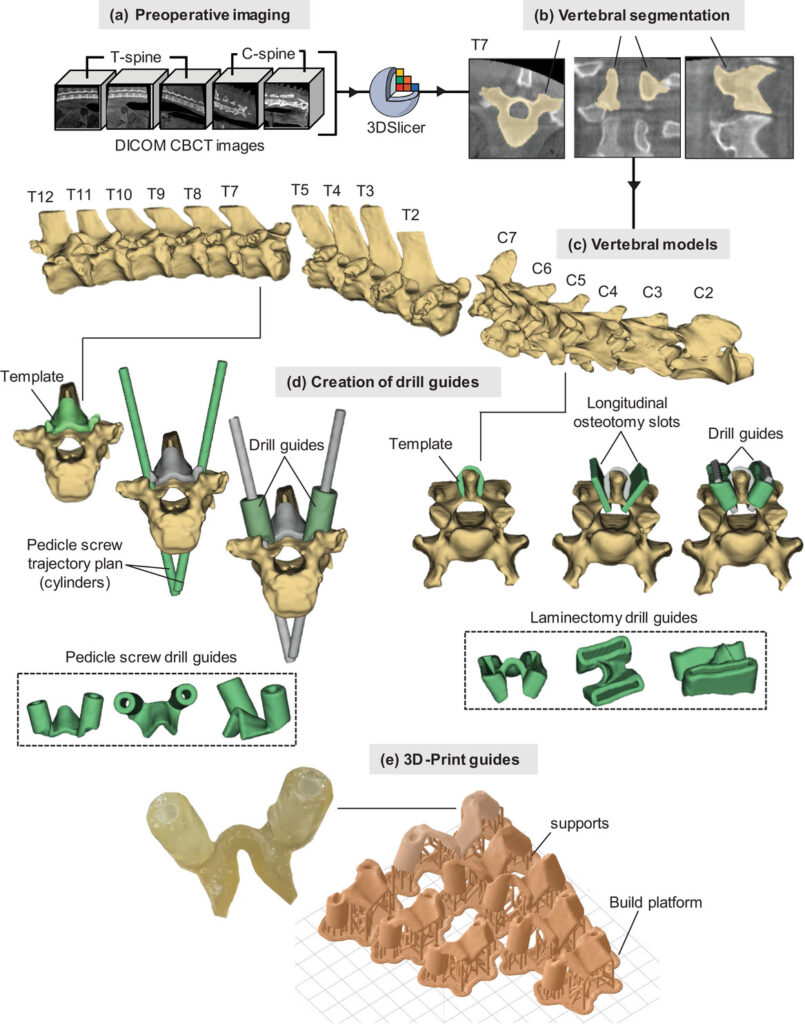
Metal hardware is often implanted to help stabilise anatomy following injury from trauma, degenerative diseases, and cancer. Accurate placement of the hardware is paramount, with misplacement potential leading to severe complications. To ensure accurate placement, 3D-printed surgical guides that allow pre-planning of the hardware position, taking into consideration individual anatomical characteristics are of growing interest. To date, 3D-printed surgical guides have been predominately developed from preoperative CT and MRI scans.
Working in collaboration with Dr Andrew Kanawati (Westmead Hospital, Sydney), Image X Interventional Imaging has been investigating the possibility of using robotic CBCT imaging system to create 3D-printed surgical guides. Robotic CBCT systems are becoming common place in orthopaedic departments, facilitating diagnosis and treatment planning in the extremities (i.e. wrist, ankle, and knee) and are ripe for expansion to other sites.
To date, we have examined the accuracy of developing 3D-printed surgical guides for spine stabilisation and laminectomy procedures. In animal cadaver models, the 3D-printed surgical guides demonstrated sufficient precision compared to those previously reported from CT.

Metal Artifact Reduction
Developing strategies to complement surgeon experience, such as utilising 3D-printed surgical guides, is a key step in reducing the risk of interventional procedures. Another step is enabling direct surgical verification of the procedure in the operating room. However, a current challenge to this is that the metal hardware itself negatively interferes with standard intraoperative 3D x-ray imaging techniques, creating unclear and imprecise images, reducing the accuracy of the assessment.
Working in collaboration with Johns Hopkins University, we have developed new non-circular CBCT imaging techniques that avoid directly imaging the hardware, dramatically improving the image quality, enabling direct in-room surgical verification of procedures.
Collaborators
Johns Hopkins University
The Advanced Imaging Algorithms and Instrumental Laboratory
Associate Professor J. Webster Stayman
Research Assistant Professor Grace J. Gang
Yiqun (Quinn) Ma (PhD student)
Johns Hopkins University
Dr Nicholas Theodore (MD) – Neurosurgery
Dr Clifford Weiss (MD) – Interventional Radiology
Westmead Hospital
Dr. Andrew Kanawati (MD) – Orthopaedics
Royal Prince Alfred Hospital
Dr Alex Constantinidis (MD)
Austrian Center for Medical Innovation and Technology
Research Center for Medical Image Analysis and Artificial Intelligence (MIAAI)
Department of Medicine, Danube Private University
Dr Sepideh Hatamikia
Industry Partners
Siemens Healthineers
OSSIS Orthopaedic Implants
Related Publications
- Kanawati, A. Constantinidis, Z. Williams, R. O’Brien, and T. Reynolds, “Generating patient-matched 3D-printed pedicle screw and laminectomy drill guides from Cone Beam CT images: studies in ovine and porcine cadavers”, Medical Physics 49 (7), 4642-4652, 2022.
- Reynolds, Y. Ma, A. Kanawati, A. Constantinidis, Z. Williams, G. Gang, O. Dillon, T. Russ, W. Wang, T. Ehtiati, C. Weiss, N. Theodore, J.H. Siewerdsen, J.W. Stayman, and R.T. O’Brien, “Extended longitudinal 3D imaging with a multi-turn reverse helical CBCT scan”, Investigative Radiology 2022.
- Y.Q. Ma, T. Reynolds, T. Ehtiati, C.R. Weiss, K. Hong, N. Theodore, G.J. Gang, J. W. Stayman, “Fully automatic online geometric calibration for non-circular cone-beam CT orbits using fiducials with unknown placement”, Medical Physics, 51 (5), 3245-3264, 2024.
- T. Reynolds, S. Hatamikia, Y. Ma, O. Dillon, G. Gang, J. Stayman, and R. O’Brien, “Technical Note: Extended longitudinal and lateral 3D imaging with a continuous dual-isocenter CBCT scan”, Medical Physics, 50 (40), 2372-2379, 2023.
- S. Hatamikia, A. Biguri, G. Herl, G. Kronreif, T . Reynolds, J. Kettenbach, T. Russ, A. Tersol, A. Maier, M. Figl, J. H. Siewerdsen and W. Birkfellner, “Source-detector trajectory optimization in cone-beam computed tomography: A comprehensive review on today’s state-of-the-art”, Physics in Medicine and Biology, 67 (16), 2022.
Invited Presentations
- Reynolds, “Integrating interventional CBCT imaging into the treatment workflow of traumatic musculoskeletal injuries” – American Association of Physicists in Medicine Annual Meeting, Washington, DC, USA (July 2022).
- Reynolds, “Non-circular trajectories in cone-beam CT imaging” – American Association of Physicists in Medicine Annual Meeting, Columbus, Ohio, USA (July 2021).
Awards
- 2022 Eureka Prize for Outstanding Early Career Researcher: Dr Tess Reynolds.
- 2021 Jack Fowler Early-Career Investigator Competition Winner: Dr Tess Reynolds, American Association of Physicists in Medicine Annual Meeting: T. Reynolds, Q. Ma, G. Gang, O. Dillon, T. Russ, W. Wang, T. Ehtiati, C. Weiss, N. Theodore, J. Siewerdsen, R. O’Brien, and J. W. Stayman, “Imaging from the lumbar to the cervical spine with a continuous multi-turn reverse helical cone-beam CT scan” – American Association of Physicists in Medicine Annual Meeting, Columbus, Ohio, USA (July 2021).
- 2021 John R Cameron Symposium Winner: Yiqun Q. Ma, American Association of Physicists in Medicine Annual Meeting: Q. Ma, T. Reynolds, G. Gang, O. Dillon, T. Russ, W. Wang, T. Ehtiati, C. Weiss, N. Theodore, J. Siewerdsen, R. O’Brien, and J. W. Stayman, “Non-circular orbits on a clinical robotic C-arm for reducing metal artifacts in orthopaedic interventions”– American Association of Physicists in Medicine Annual Meeting, Columbus, Ohio, USA (July 2021).
In the Media
- Television interview with Kathryn Robinson: ABC Mornings (Nationwide), aired 11:27am September 1, 2022.
- Radio interview with Josh Szeps on ABC Sydney, aired 2:30pm September 1, 2022.
Eureka Prizes 2022: Outstanding Early Career Researcher Prize Winner Dr Tess Reynolds: https://www.youtube.com/embed/osyuuk4voU0
University of Sydney news article: Pioneers recognised in Australia’s top science prizes
https://www.sydney.edu.au/news-opinion/news/2022/09/01/pioneers-recognised-in-australia-s-top-science-prizes.html


