
Reducing the impact of radiation on the heart for cancer and cardiac disease patients
The Cardiac Radioablation program is funded by an NHMRC Synergy Grant.
Background
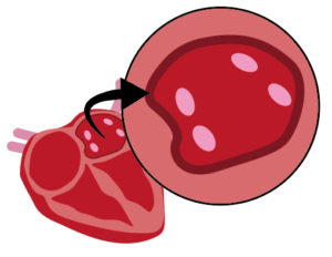
Ventricular tachycardia (VT) and atrial fibrillation (AT) are cardiac conditions that affect over 33.5 million people globally every year. Treatment typically involves invasive heart surgery to manually create scar tissue in specific areas of the heart, to block the abnormal heart signals. Radiation therapy is emerging as a promising new non-invasive treatment option.
Cardiac radioablation is a revolutionary non-invasive method of producing cardiac scarring. The potential of radioablation has been demonstrated through recent clinical trials, however there is considerable room to improve this emerging treatment. To ensure cardiac radioablation treatment efficacy and safety, it is vital that radiation is carefully delivered to the cardiac target, but steered away from the healthy organs and cardiac substructures that are often in very close proximity. A considerable challenge is that the heart is moving with both cardiac contraction and respiration. We are addressing this motion management problem for cardiac radioablation by inventing imaging technologies for the entire treatment pathway from CT simulation, to pre-treatment patient-connected imaging, and during treatment delivery.
Team Contact
For more information about the program and projects, contact Dr Mark Gardner

Expressions of Interest
If you’re seeking a new challenge, Image X welcomes expressions of interest from researchers with expertise in fields including algorithm development, machine learning, advanced hardware use, and clinical translation across all imaging modalities are welcomed.
Team
| Investigator | Research Institution |
|---|---|
| Prof Ricky O’Brien | RMIT University |
| Prof Paul Keall | Image X Institute – University of Sydney |
| Dr Doan Trang Nguyen | Image X Institute, University of Sydney |
| Assoc Prof Shankar Siva | Peter MacCallum Cancer Centre |
| Dr Tess Reynolds | Image X Institute, University of Sydney |
| Assoc Prof Saurabh Kumar | Western Sydney Local Health District |
| Assoc Prof Lois Holloway | University of New South Wales |
Project Collaborations
Through a wide array of projects, we’re collaborating with experts across multiple research institutes, clinical sites, and imaging vendors to unleash the full potential of cardiac radioablation.
Accurate CT imaging in the presence of respiratory and cardiac motion (Keall, O’Brien)
In a successful collaboration with Siemens, we’ve countered motion artefacts in images, caused by the patients breathing. Building on this, we’re integrating cardiac motion by adjusting CT table motion and image capture based on real-time respiratory and cardiac signals. This technology can also predict errors from respiratory and cardiac data, pausing the CT beam during anomalies for accurate imaging.
Using an MRI-Linac for real time cardiac tracking and substructure avoidance (Holloway, Keall)
This project expands on our technology to track and monitor the motion of the ablation target in real-time during treatment. The work takes advantage of the exquisite image quality made available by MRI scanners. This research is being conducted in collaboration with the Australian MRI Linac Program, on a novel MRI-Linac (one of only 4 research systems in the world) located at Liverpool Hospital.
Cardiac and respiratory imaging on the treatment table (O’Brien, Reynolds)
Utilising 4DCBCT technology, in this project patients will be positioned for treatment with time-resolved CT images, captured during breathing. Sequential display of these images aids treatment alignment to the planned area, and shows clinicians when re-adjusting is needed. Validation will involve cardiac phantom tests to assess imaging precision.