Leadership
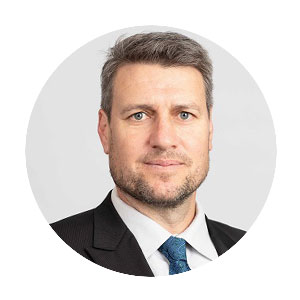
Professor Paul Keall
Director
Professor Paul Keall is the Director of the Image X Institute. He and his team improve lives by inventing and advancing new ways to image and treat disease through new technologies for medical imaging and targeted radiation therapy. Specific technologies include real-time image and dose-guided radiation therapy, adaptive radiation therapy, and functional imaging, His work benefits patients by ensuring high doses of radiation are targeted at the cancer to improve the chance of cure; and simultaneously that high doses of radiation avoid healthy tissue to reduce the chance of treatment side effects that will impact patients’ quality of life. He works with researchers, clinicians, patients, and industry to create a pipeline of innovations that are translated and accessible to people with cancer worldwide.
Prof. Keall’s research is funded by over $8M of competitive government grant funding. The scientific work has resulted in over 400 articles (h-index 90). The impact of his research is evidenced by founding three companies, inventing 40 patents, and completing 20 intellectual property agreements with ten companies that have to date resulted in five globally available products.
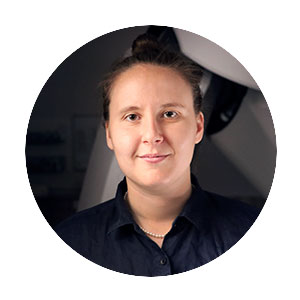
Associate Professor Tess Reynolds
Deputy Director
Associate Professor Tess Reynolds is an NHMRC Emerging Leadership (EL2) and Robinson Fellow at the University of Sydney. Her international academic-industry-healthcare research programs are focused on unlocking the full potential of robotic imaging systems to deliver the next generation of intra-operative and interventional imaging. In 2022, she won an Australian Museum Eureka Prize (Outstanding Early Career Researcher) and has secured $9.6M in competitive grant funding to date.
A/Prof Reynolds has a passion for leadership and mentoring. In 2024 A/Prof Reynolds was named an “Emerging Leader of Academic Medical Physics” by the University of Wisconsin-Madison (USA) and served as the inaugural chair of the Image X Mentoring Committee, overseeing a structured mentoring program for the Institute. Dr Reynolds also holds an appointment to the Leadership Group of the University of Sydney’s Sydney Clinical Imaging Network (SCIN), convening the 2025 SCIN Research Summit, and serves as a member to the Image Physics Committee of the American Association of Physicists in Medicine. At the Institute, A/Prof Reynolds curates and hosts a month-long public ‘Emerging Women in Medical Physics and Radiology’ seminar series in November of each year.
Academic Staff
Dr Thomas Boele
Research Associate | View academic profile
Dr Thomas Boele designs, builds and deploys prototype devices at the intersection between quantum physics and medical imaging. He is currently developing accessible MRI techniques to image metabolic changes associated with cancer for better diagnosis and treatment guidance.
Thomas collaborates with researchers at Harvard/MGH, Wayne State and NCSU to advance low-field hyperpolarized MRI technologies. He welcomes student and collaborator interest in parahydrogen-based hyperpolarization and innovation in MRI hardware.
Dr Vicky Chin

Research Associate
Project: Cardiac Radioablation
Dr Vicky Chin is a radiation oncologist at Liverpool and Macarthur Cancer Therapy Centres. Her PhD examined the cardiac effects of radiotherapy for patients who have received treatment for thoracic cancers. She is part of the NHMRC Synergy Grant research team looking to better understand cardiac responses to radiation, to improve treatments for both for cancer and cardiac patients.
Dr Owen Dillon

Research Fellow | View academic profile
Project: The Patient Connected Imaging Program
Dr Owen Dillon focuses on Cone Beam Computed Tomography (CBCT) with a particular emphasis on estimating and incorporating patient motion. He is primarily involved in the ADAPT clinical trial, improving lung cancer radiotherapy imaging while reducing imaging time 60% and imaging radiation 85%.
Dr Dillon’s research also involves robotic systems in surgical theatres and implementing 3D imaging capabilities on 2D systems. His background is in Image Reconstruction, Inverse Problems and Bayesian Statistics.
Dr Mark Gardner

Research Fellow | View academic profile
Project: Cardiac Radioablation
Dr Mark Gardner is using deep learning and advanced image processing methods to monitor and compensate for different types of patient motion during radiation therapy. Dr Gardner’s work on monitoring patient motion aims to improve the accuracy of cardiac radioablation treatments and to provide faster and more accurate lung cancer treatments in fewer treatment session. He is also working to reduce patient anxiety in head and neck cancer patients during radiation therapy treatments by removing the need for immobilisation masks. Dr Gardner has partnered with Royal North Shore Hospital, Westmead Hospital and Blacktown Hospital to bring his innovations to patients in the clinic.
Dr Gardner uses deep learning-based segmentation and registration methods, as well as cone-beam CT reconstruction methods to achieve his research goals. Dr Gardner aims to implement real-time tracking of key organs during radiation therapy including head and neck tumours and cardiac substructures. He is also looking at developing methods for simulation-free single fraction lung cancer radiation therapy treatments. He welcomes expressions of interest from students and collaborators looking for challenges in image processing and CT reconstruction methods and to improve radiation therapy treatments.
Dr Emily Hewson
Cancer Institute NSW Early Career Research Fellow | View academic profile
Project: Kilovoltage Intrafraction Monitoring (KIM)
Dr Emily Hewson develops motion monitoring and tracking techniques for radiation therapy She received her PhD from the University of Sydney in 2022; her thesis focused on developing beam-adaption that can track multiple targets simultaneously for patients with locally advanced cancer and was carried out in collaboration with the Royal North Shore Hospital. Dr Hewson’s work in this field was awarded Best in Physics in 2021 at the 63rd AAPM annual meeting.
Dr Nicholas Hindley

Cancer Institute NSW Early Career Research Fellow | View academic profile
Project: Markerless Tracking
Dr Nicholas Hindley is a Cancer Institute NSW Early Career Fellow and Honorary Fellow of the National Heart Foundation. He leads a research program centred around AI-guided cancer care and developed the open-source VoxelMap deep learning framework for tumour and organ tracking during radiotherapy.
To drive both technology development and clinical translation, Dr Hindley has partnered with UCLA, Duke University, the Health Translation Hub at UNSW and the Royal North Shore and Royal Prince Alfred hospitals. With his work, Dr Hindley hopes to improve both the cure rate and quality of life of people living with cancer. In furthering this mission, he is always looking to work with passionate, enthusiastic and self-driven researchers and students.
Dr Andrew Phair

Research Associate | View academic profile
Project: The Australian MRI-Linac Program
Dr Andrew Phair leverages artificial intelligence techniques to improved real-time tumour tracking in MR-guided radiotherapy. He received his PhD in applied mathematics from the University of Tasmania in 2020 and subsequently took up a postdoctoral position at the School of Biomedical Engineering and Imaging Sciences at King’s College London, where his work focused on deep-learning techniques to accelerate and increase the achievable resolution in whole-heart cardiac MRI.
Dr Chandrima Sengupta

Cancer Institute NSW Early Career Research Fellow | View academic profile
Project: Kilovoltage Intrafraction Monitoring (KIM)
Dr Chandrima Sengupta works to develop, improve and advance real-time adaptive radiotherapy techniques for radiation therapy patients. She has led the clinical implementation of Kilovoltage Intrafraction Monitoring (KIM) technology in two multi-institutional clinical trials: TROG 17.03 LARK trial and TROG 18.01 NINJA trial that aim to quantify the geometric and dosimetric accuracy achieved with KIM guidance for liver and prostate cancer patients. She collaborates on the Remove the Mask project, on testing the accuracy of novel surface guidance applications using a 6 degree-of-freedom quality assurance device she co-authored and manages. She is also interested in imaging technologies related to proton therapy treatment.
Dr David Waddington

NHMRC Emerging Leadership Fellow | View academic profile
Project: The Australian MRI-Linac Program
Dr David Waddington leads the MRI research program at the Image X Institute, where he develops advanced imaging technologies to improve cancer diagnosis and treatment. His work focuses on creating better ways to see the body, so treatments can adapt precisely to changes in tumour biology and anatomy.
David holds competitive fellowships and grants, including an NHMRC Investigator Grant, and collaborates internationally, including with Harvard Medical School. He welcomes students and collaborators interested in translating next-generation MRI technologies that integrate low-field MRI, MRI-guided radiotherapy and artificial intelligence.
Ann Yan

Research Associate
Project: Kilovoltage Intrafraction Monitoring (KIM)
Ann Yan is developing hardware, software and machine learning algorithms to advance image-guided radiotherapy. Ann completed her Bachelor of Engineering Honours (Biomedical) in 2021 and Master of Data Science in 2022 at the University of Sydney. Ann has worked as a biomedical engineer in Prince of Wales hospital along with Royal Women’s Hospital and Sydney Children‘s Hospital, specialized in pacemaker implantation and troubleshooting.
Dr Zhuang Xiong

Research Associate | View academic profile
Project: Kilovoltage Intrafraction Monitoring (KIM)
Dr Zhuang Xiong develops AI-driven, marker-less tumour tracking methods to improve dose delivery precision for real-time image-guided radiotherapy. His current work investigates 2D prompt-based and foundation segmentation models, and 3D image reconstruction techniques for marker-less tumour tracking. These approaches aim to improve treatment accuracy and reduce patient burden by eliminating implanted markers.
Zhuang collaborates across medical physics, AI, and clinical disciplines to translate innovative methods into reliable tools for adaptive radiotherapy.
Research Support Staff
Dr Helen Ball

Operations Manager | Contact
Dr Helen Ball manages the operations of Image X Institute, ensuring that the institute’s research programs run smoothly and efficiently. Helen has a longstanding interest in research support, informed by her previous experience as a researcher at the University of Sydney. She is dedicated to enabling impactful research and improving systems and processes that help research teams thrive.
Alen Biju

Research Assistant
Project: Interventional Imaging
Alen Biju is developing metal-artifact reduction imaging for CBCT-guided bronchoscopic lung biopsies. He completed his Master of Medical Physics at the University of Sydney , with a thesis focused on developing a single-projection CBCT method for the real-time localisation of HDR brachytherapy sources.
Michael Ferraro

Research Assistant
Project: The Australian MRI-Linac Program
Michael Ferraro works on MRI reconstruction techniques for speech imaging and low-field MRI. He is also contributing to a project developing parahydrogen-based hyperpolarization for MR-guided radiotherapy. He recently completed his Master of Medical Physics and has a strong interest in MRI research and its applications.
Rose Hodges

Research Assistant
Project: The Australian MRI-Linac Program
Rose Hodges is developing low-field MRI technologies, including commissioning an MRI spectrometer with a Halbach array and creating new probes based on hyperpolarisation for imaging brain cancer. She completed her Master of Physics at the University of Manchester in May 2024. For her final project, she worked in a small team to build a low-cost Earth’s Field NMR spectrometer and design a spin-echo sequence for it.
Freeman Jin

Software Engineer
Project: Kilovoltage Intrafraction Monitoring (KIM)
Freeman Jin is a software engineer working on the implementation of Megavoltage imaging segmentation on KIM. A passionate “imaging addict” both inside and outside the office, Freeman devotes most of his time to exploring all aspects of imaging – from everyday photography to astrophotography and advanced image processing. His enthusiasm for capturing and analysing visual data has now found its professional home in the field of medical imaging.
Julia Johnson
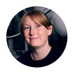
Design & Communications Officer | Contact
Julia Johnson provides a full suite of design services for the institute including design and fabrication of class 1 medical device prototypes, as well as graphics, branding, scientific figures, and content creation. In addition to this, Julia is passionate about communicating the work of the institute to the public. She manages the website and social media channels, and strengthens the institute’s engagement with the wider community.
Yu (Sam) Liang

Software Engineer
Project: Kilovoltage Intrafraction Monitoring (KIM)
Sam Liang works on developing databases to enable our medical imaging research, He has a background in engineering and he completed his Master of Information Technology at UNSW. He has also worked as a software engineer in the aerospace industry, gaining valuable experience in developing scientific software solutions for space missions.
Dr Marcel Schulz

Clinical Trials Co-ordinator | Contact
Dr Marcel Schulz coordinates the VITaL healthy lung sparing trial. The CT Ventilation Imaging technique being examined in this trial is a novel software-based method developed by Image X that maps functional lung regions using standard pre-treatment CT scans acquired during breath-hold inhalation and exhalation phases.
At the NHMRC CTC, Marcel also supports a project investigating how presentation considerations affect the administration of Patient Reported Outcome Measures to people with chronic health conditions. Marcel’s research interests include evaluating the health-related quality of life among marginalized populations, such as individuals with chronic health conditions and people who inject drugs.
Shona Silvester
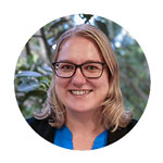
Clinical Trials Lead | Contact
Shona Silvester supports the clinical trials program at the Image X Institute, contributing across protocol development, site start-up, data management, and trial coordination. Her work plays a vital role in translating innovative research from bench to bedside, enabling patients to access cutting-edge technologies and treatments through interventional clinical trials.
With extensive experience in clinical trials research, particularly in oncology, Shona brings expertise and a strong commitment to advancing patient care through rigorous and impactful clinical studies.
Emma Wisser

Intern – Science Communication and Illustration
Emma is a University of Newcastle student studying Microbiology and Illustration. She has joined the Image X Institute on placement, where she is excited to gain experience as a science communicator and designer under the supervision of Julia Johnson.
Higher Degree Research Students
Chen Cheng
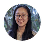
PhD Candidate
Chen Cheng investigates real-time monitoring techniques for head and neck cancer radiotherapy using x-ray and surface imaging. She graduated with a Bachelor of Science and Bachelor of Advanced Studies at the University of Sydney in 2022, majoring in mathematics and physics. Previously during her undergraduate studies, she interned at Image X, assisting with the implementation of a surface guidance system for the Remove the Mask project.
James Grover
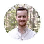
PhD Candidate
James Grover explores how deep learning can be used to better treat and diagnose disease. He’s applying artificial intelligence algorithms to MRI-guided radiation therapy to enable more accurate treatment delivery and boost the diagnostic potential of portable ultra-low-field MRI. James’ interest in inventing and applying novel algorithms stems from a desire to bridge the gap between cutting-edge computational methods and real-world clinical impact.
Jonathan Hindmarsh

PhD Candidate | Contact
Jonathan Hindmarsh is investigating the implementation of real time treatment techniques. Jonathan has worked as a Medical Physics Specialist in NSW, Australia and Alberta, Canada since getting his qualification in 2013 and his research interests include the development and improvement of treatment techniques, commissioning methods, and quality assurance processes.
Alicja Kaczynska
PhD Candidate
Project: Kilovoltage Intrafraction Monitoring (KIM)
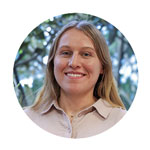
Alicja Kaczynska is working to develop image-guided radiotherapy technology to improve motion management for abdominal and thoracic radiotherapy patients. Her research combines x-ray and surface imaging through a correlation model for real-time respiratory motion monitoring on standard treatment machines. Alicja previously completed a Master of Medical Physics validating a motion correlation model on a liver SBRT dataset from Image X Institute’s TROG LARK clinical trial.
Alicja is committed to providing opportunities for others interested in a STEM pathway – each year she co-ordinates symposia for HDR students interested in STEM research.
Kaan Kandasamy

PhD Candidate
Project: Kilovoltage Intrafraction Monitoring (KIM)
Kaan Kandasamy in working on clinical validation and implementation of AI based real-time tumour tracking for radiation therapy. Kaan is a clinical medical physicist at GenesisCare and is the lead physicist for the PRIME clinical trial for implementing the KIM marker tracking technology on Elekta linacs across the county.
Jeremy Lim

PhD Candidate
Project: CT Ventilation Imaging
Jeremy Lim is a PhD candidate using non-contrast CT imaging to map airflow (ventilation) and blood flow (perfusion) in the lungs. His research aims to make lung cancer radiation therapy safer by tailoring treatment to each patient’s lung function and sparing healthy lung regions. This approach has the potential of reducing the risk of radiation-induced lung toxicity such as pneumonitis, and improving post-treatment quality of life.
Gregory Willson

PhD Candidate
Project: Kilovoltage Intrafraction Monitoring (KIM)
Greg Willson is working on markerless liver tumour tracking for radiotherapy using AI-based methods. He completed a Bachelor of Science and Bachelor of Advanced Studies at the University of Sydney in 2024, majoring in Physics and Mathematics. For his honours project in physics, he investigated Positron Lifetime Spectroscopy techniques.
